Research Symposium
23rd annual Undergraduate Research Symposium, April 6, 2023
Makenna Jenkins Poster Session 3: 2:45 pm - 3:45 pm/ Poster #243

BIO
My name is Makenna Jenkins and I am a first year from Lehigh Acres, Florida studying Cell and Molecular Neuroscience. I joined the Gratton Lab during August of 2022 and have loved every moment of my time in the lab. My research interests include physiological data, migraines (heritability, neurological locations and differences, and treatments), genetic applications to neuroscience, and the medical applications of hallucinogens (as well as many others). I plan to pursue a higher educational degree after undergrad, and am currently deciding between Medical School and Graduate School (or both). Along the lines of my future goals, I intend to continue my research career and possibly become a PI of my own lab.
Physiological Data and fMRI
Authors: Makenna Jenkins, Caterina GrattonStudent Major: Cell and Molecular Neuroscience
Mentor: Caterina Gratton
Mentor's Department: Neuroscience Mentor's College: Undergrad- University of Illinois; Grad School- UC Berkeley; Currently- Florida State University Co-Presenters:
Abstract
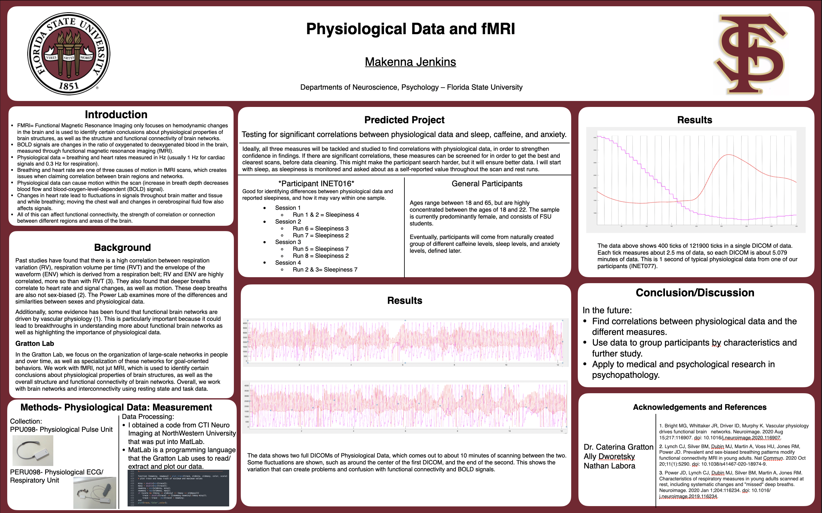
Functional Magnetic Resonance Imaging (fMRI) is currently one of the best ways for researchers, scientists, and doctors to view and map the brain. Different from MRI, fMRI measures changes in the ratio of oxygenated to deoxygenated blood in the brain, which gives us the blood-oxygen-level-dependent (BOLD) signal. Understanding how heart rate and respiration (physiological) data affects fMRI is imperative not only to the researcher, but to anyone who follows their research. BOLD signals are often noisy, meaning the images aren’t clear due to motion, systemic complications, and physiological alterations. A measure as simple as breathing can introduce more noise into the BOLD signal in fMRI, but is usually glossed over even though it has been identified as one of three main reasons for noisiness. Specifically, breaths can cause motion in brain images, largely impacting data quality; it even alters the results of Functional Connectivity analysis, a measure of how strongly connected parts of the brain are at rest or during tasks. Physiological data is collected through scanner tools such as a pulse oximeter and respiratory belt, then generated through different steps of coding applications like MatLab. This turns into a full scale image of every breath taken, and heart rate, throughout the scan. Physiological data’s effect is commonly disregarded by many cognitive psychologists, which is why this presentation will discuss the importance of collecting and analyzing this data, as well as how to potentially apply it to individuals for measure comparisons (amount of sleep, anxiety/stress levels, caffeine intake).
Keywords: Physiological data, fMRI


