Research Symposium
25th annual Undergraduate Research Symposium, April 1, 2025
Elle Plasencia Poster Session 3: 1:45 pm - 2:45 pm/ Poster #83

BIO
Elle Plasencia is a sophomore from Stuart, Florida studying Art History with a minor in Museum Studies. She is also in the FSU Honors Program and Undergraduate Research Opportunity Program (UROP). Outside of her classes, she spends her time working in the Special Collections & Archives and rehearsing with Campus Band. After graduation, she aspires to go into an art history, conservation, or preservation graduate education or career field. Her UROP project involves using various microscopy techniques to create artistic photographs of microorganisms, and her further research interests include the relationships between art and science and the evolution of art mediums and production over time.
Microscopic BioArt
Authors: Elle Plasencia, Jamel AliStudent Major: Art History
Mentor: Jamel Ali
Mentor's Department: Department of Chemical and Biomedical Engineering Mentor's College: FAMU-FSU College of Engineering Co-Presenters:
Abstract
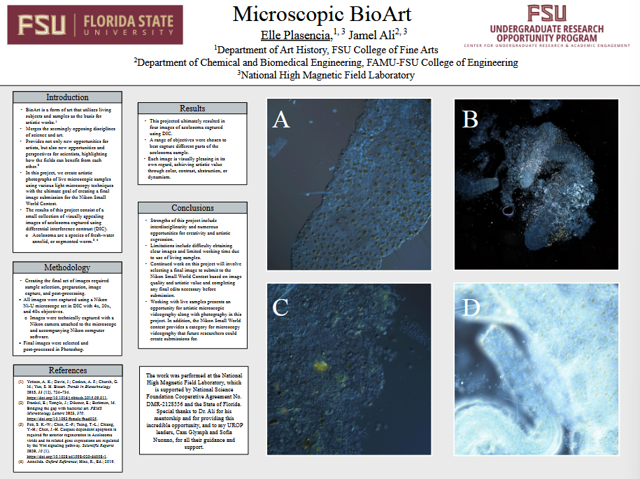
In this project we create artistic photographs of live microscopic samples using various light microscopy techniques with the ultimate goal of submitting a final image to the 2025 Nikon Small World contest. Microscopy and microscopic samples are rarely the subject of art, and are more often utilized by scientists to investigate the microscopic world. However, microscopy can be used to open the door for new forms of artistic expression. With light microscopes, the macroscopic world is no longer the limit for what can be visually captured with traditional photography. Here, creating photographs first required sample preparation, involving concentrating live samples in a centrifuge and carefully pipetting them into glass slide chambers. Then, various widefield techniques were utilized to generate raw images, including brightfield, darkfield, and differential interference contrast (DIC). Once images were obtained, post-processing took place in editing programs including DaVinci Resolve and Photoshop. The result is a small collection of visually pleasing images of microscopic invertebrates in DIC. Some images are of singular organisms, taken from very close working distances (<1mm), while others feature multiple organisms within one field of view. The Microscopic BioArt presented here solidifies the existing connection between science and art as disciplines and broadens horizons of possibility for both scientists and artists.
Keywords: microscopy, microorganisms, bioart, photography


